Digital biomechanics
AI and multiscale mechanics for a gradient 3D-printed joint implant
Gait data, a cellular mechanical window and combined DLP/inkjet printing are linked into a programmable PEGDA/beta-TCP gradient.
Authorized republication with translated text and the source’s original scientific figures. Numerical results belong to the reported study, not to KUNWU equipment specifications. This is laboratory or preclinical research, not a clinical recommendation or evidence of a medical device’s clinical efficacy.
- Stress-concentration factor
- ≤1.23
- Compliance-match coverage
- 88.5–93.7%
- Reconstruction error
- 3.8–5.1 kPa
The source describes a graded multimaterial 3D-printing approach guided by multiscale biomechanical modeling. By modeling mechanical transfer from macroscopic movement to microscopic cellular responses, the study aims to fabricate biomimetic joint implants integrating structural, functional and topological gradients.
1. Research background
More than one million joint-replacement procedures are performed worldwide annually, according to the source. Despite successful replacement, poor integration between the implant and host tissue remains a long-term clinical and engineering challenge.
Conventional metal-alloy or ceramic implants are foreign structures whose design often emphasizes replacing damaged tissue with a stronger material. A human joint, however, is not a simple mechanism; it is a coupled mechanical system operating across multiple scales.
During walking, articular cartilage experiences dynamic loads of approximately three to five times body weight. Load passes from a soft surface layer with an elastic modulus around 0.5–2 MPa, through calcified cartilage at approximately 100–300 MPa, into subchondral bone at roughly 1–10 GPa. This soft-to-hard gradient is a central feature of natural joints.
Homogeneous implants may be too stiff or too compliant relative to adjacent tissue. Mechanical mismatch concentrates stress at the interface, repeatedly loading vulnerable new tissue and potentially contributing to loosening, displacement and revision surgery.
2. Research approach
The central concept is to treat implant design not solely as a structural engineering problem but also as a problem of cellular mechanotransduction.
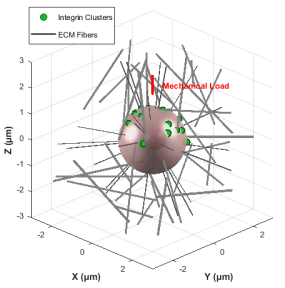
Cells continuously sense extracellular-matrix mechanics through molecular structures such as integrins and focal adhesions. Pathways including YAP/TAZ influence proliferation, differentiation and apoptosis. The source describes a mechanical window compatible with cellular homeostasis: stimulation above or below that window may disrupt cell function.
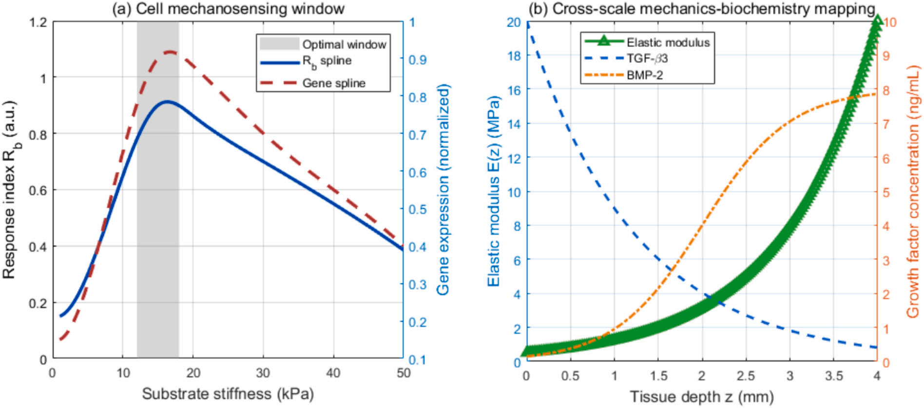
The team used real gait data in a multiscale finite-element model to examine stress transmission through cartilage, the calcified layer and subchondral bone. Atomic force microscopy and analysis of YAP/TAZ signaling were used to infer mechanical ranges associated with cellular homeostasis, including modulus and dynamic-strain amplitude. These parameters provided biological constraints for material design.
The next task was to convert the quantified mechanical window into a manufacturable architecture.
The proposed coupled-gradient strategy combined an exponential elastic-modulus gradient, approximating the cartilage-to-bone transition, with gradients in BMP-2 and TGF-beta to guide region-specific chondrogenic or osteogenic differentiation.
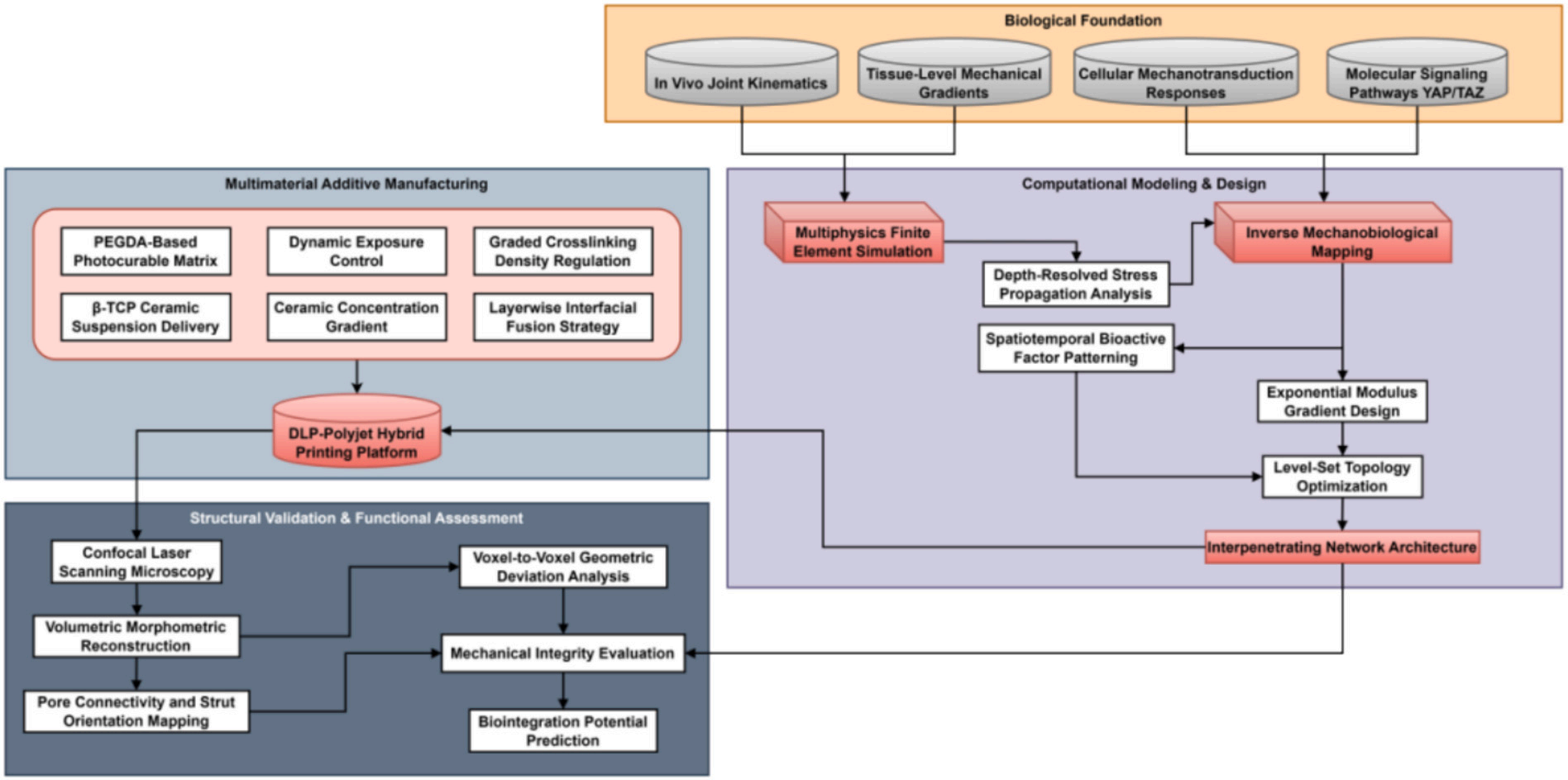
A graded multimaterial process combined digital light processing with inkjet deposition:
DLP
rapidly cured PEGDA resin to provide structural support.
Inkjet printing
precisely deposited droplets containing beta-TCP ceramic particles. Spatial control of exposure time and beta-TCP concentration produced a continuous mechanical gradient.
A unified G-code schedule synchronized DLP projection patterns with inkjet deposition in space and time. Droplets were deposited before the resin reached its gel threshold, aiming to avoid phase separation and maintain a uniform component distribution.
A level-set method also optimized interface topology to create interpenetrating networks, limit stress concentration and improve mechanical continuity and biological compatibility.
3. Reported results
Mechanical performance: the reported average gradient smoothness was at least 4.59 × 10^3 micrometres. Under two loading conditions, the mean stress-concentration factor did not exceed 1.23, outperforming the compared homogeneous and simple multilayer designs.
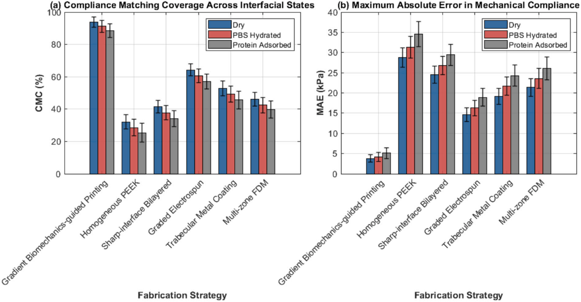
Cell-microenvironment reconstruction: across physiologically relevant interface conditions, the method showed mean compliance-matching coverage of 88.5–93.7%. Mean reconstruction error ranges for local mechanical parameters were 3.8–5.1 kPa.
Confocal imaging was used to assess geometry and composition distribution. The source reports satisfactory accuracy and uniformity, supporting the manufacture of structures that combine biomimetic geometry and functional gradients.
4. Conclusions
The broader significance proposed in the source is a shift from passive support toward designs that guide tissue regeneration, rather than merely addressing the immediate structural requirements of an implant.
The source contrasts a conventional implant as a temporary functional support with a biomimetic scaffold intended to direct cell growth and support integration with host tissue. This is a research concept; the reported modeling and fabrication results do not establish clinical efficacy.