Bioceramics
HAp scaffold porosity for bone regeneration: why about 400 um performed best
A four-scaffold comparison shows how porosity, pore size, wall thickness, strength and cell infiltration move together.
Authorized republication with translated text and the source’s original scientific figures. Numerical results belong to the reported study, not to KUNWU equipment specifications. This is laboratory or preclinical research, not a clinical recommendation or evidence of a medical device’s clinical efficacy.
- Porosity of the best group
- 45.61%
- Average pore size
- ~400 um
- Compressive strength
- ~1.6 MPa
More than two million bone-grafting operations are performed worldwide each year, making bone the second most commonly transplanted tissue after blood. Autografts and allografts remain reference treatments, but have limitations including donor-site injury, limited tissue supply and rejection risk. The source reports an approximately 7% annual growth rate for the global bone-graft market and a projected value of USD 4.6 billion in 2030. It links demand to population ageing—citing a WHO projection that the proportion of people aged over 60 will rise from 12% to 22%—and advances in bone tissue engineering.
Hydroxyapatite (HAp, Ca10(PO4)6(OH)2), chemically similar to the inorganic component of human bone, and faster-degrading beta-tricalcium phosphate (beta-TCP) are widely used synthetic scaffold ceramics. Both belong to the calcium-phosphate family. HAp is bioactive: it supports osteoblast-mediated bone formation and can be resorbed by osteoclasts. However, bulk HAp has limited load-bearing mechanical performance. One approach is to design porous HAp scaffolds.
What porosity and pore size allow cell infiltration without sacrificing too much strength? Published answers differ, with some studies favoring 70% porosity and others 50%. A team at Tampere University in Finland used DLP ceramic 3D printing to conduct a systematic comparison.
1. Experimental design: four porosities and five sintering temperatures
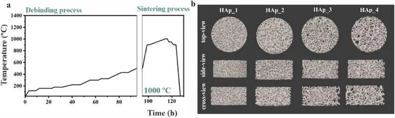
HAp slurry was printed at 45°C using a DLP printer with 25-micrometre layers. Solid discs, 7 mm in diameter, were first used to study sintering temperatures from 900°C to 1300°C, with specimens designated HAp_900 through HAp_1300. Four porous cylindrical scaffolds, 3.5 mm high and 7 mm in diameter, were designated HAp1 to HAp4 in order of increasing porosity. Micro-CT quantified their structures. All porous scaffolds were sintered at 1000°C to balance bioactivity and mechanical performance.
2. The selected balance: approximately 45% porosity and 400-micrometre pores
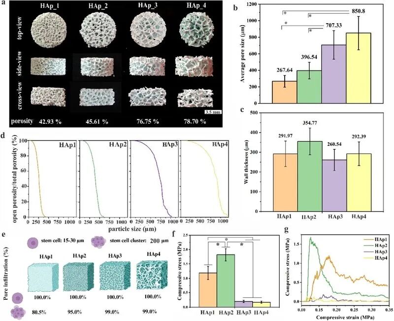
Micro-CT established the following structural parameters:
HAp1: total porosity 42.93%; average pore size 267.6 micrometres; wall thickness 292.0 micrometres; compressive strength approximately 1.2 MPa.
HAp2: total porosity 45.61%; average pore size 396.5 micrometres; wall thickness 354.8 micrometres; compressive strength approximately 1.6 MPa, the highest in the comparison.
HAp3: total porosity 76.75%; average pore size 707.3 micrometres; wall thickness 260.5 micrometres; compressive strength approximately 0.3 MPa.
HAp4: total porosity 78.70%; average pore size 850.8 micrometres; wall thickness 292.4 micrometres; compressive strength approximately 0.3 MPa.
HAp2, with 45.61% porosity and pores close to 400 micrometres, had the best mechanical performance among these designs. Smaller pores can limit cell and vessel ingrowth; larger pores of 700–850 micrometres offered up to 99% infiltration but reduced strength to about one-fifth of HAp2. Tests with 30-micrometre standard particles indicated 100% open porosity in all scaffolds, so connectivity was not the limiting issue for pores above 267 micrometres. However, individual cells of 15–30 micrometres and approximately 200-micrometre cell clusters behaved differently: cluster infiltration reached 99% for HAp3 and HAp4, but only 80.5% for HAp1. A pore size around 400 micrometres therefore provided a compromise between strength and infiltration in this experiment.
3. Biological evaluation: HAp2 showed the strongest osteogenic response
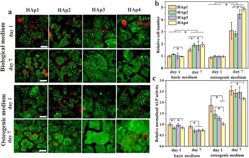
Human bone-marrow mesenchymal stromal cells (hBMSCs) from two donors were seeded onto the four scaffolds and cultured in osteogenic medium for 14 and 21 days. HAp2 showed the highest cell activity and proliferation at all measured time points. Type I collagen expression, an important osteogenic marker, was also strongest on HAp2.
The source attributes this balance to HAp2’s 45.61% porosity and approximately 400-micrometre pores, which allow clusters to penetrate, together with approximately 355-micrometre walls that provide supporting surfaces. The higher-porosity designs provide more void space but thinner walls and less attachment surface; the source describes their relatively thin walls as approximately 260 micrometres.
4. The sintering-temperature trade-off
HAp is highly bioactive but mechanically weak in its natural state. Higher sintering temperatures improve densification and strength, yet HAp has limited high-temperature stability: impurities and substituting ions can promote transformation into beta-TCP.
SEM of HAp discs sintered at 900–1300°C showed grain growth and densification. Osteoclast attachment was best at 1000°C. Above approximately 1200°C, a glassy bonding phase appeared at the surface and attachment declined as surface chemistry changed.
XRD-based phase analysis showed that deviation of the Ca/P molar ratio from the stoichiometric value of 1.67 can promote beta-TCP formation at high temperature. Biological apatite is itself calcium-deficient, and this issue is particularly important for Sr-, Mg- or Zn-substituted HAp. The team selected 1000°C for the porous scaffolds to preserve bioactivity, accepting a mechanical trade-off.
5. An unexpected result from trace-element substitution
Bone apatite accommodates a range of substituting ions. The team tested Sr-, Mg- and Zn-containing HAp formulations, designated 1MIX and 5MIX, expecting an osteogenic benefit. Instead, the substituted compositions deviated from stoichiometric HAp and formed substantial beta-TCP after sintering at 1000°C, especially in 5MIX.
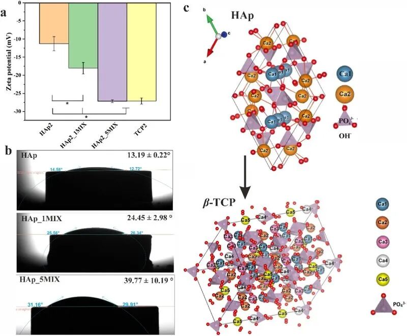
Surface measurements helped explain the result: increasing beta-TCP content was associated with a larger absolute zeta potential, meaning stronger negative surface charge, and a larger water contact angle, meaning poorer wettability. The combination created less favorable electrostatic and hydration conditions for attachment. Quantification of tartrate-resistant acid phosphatase (TRAcP), an osteoclast marker, confirmed markedly fewer osteoclasts on MIX specimens than on pure HAp2.
6. Conclusions and outlook
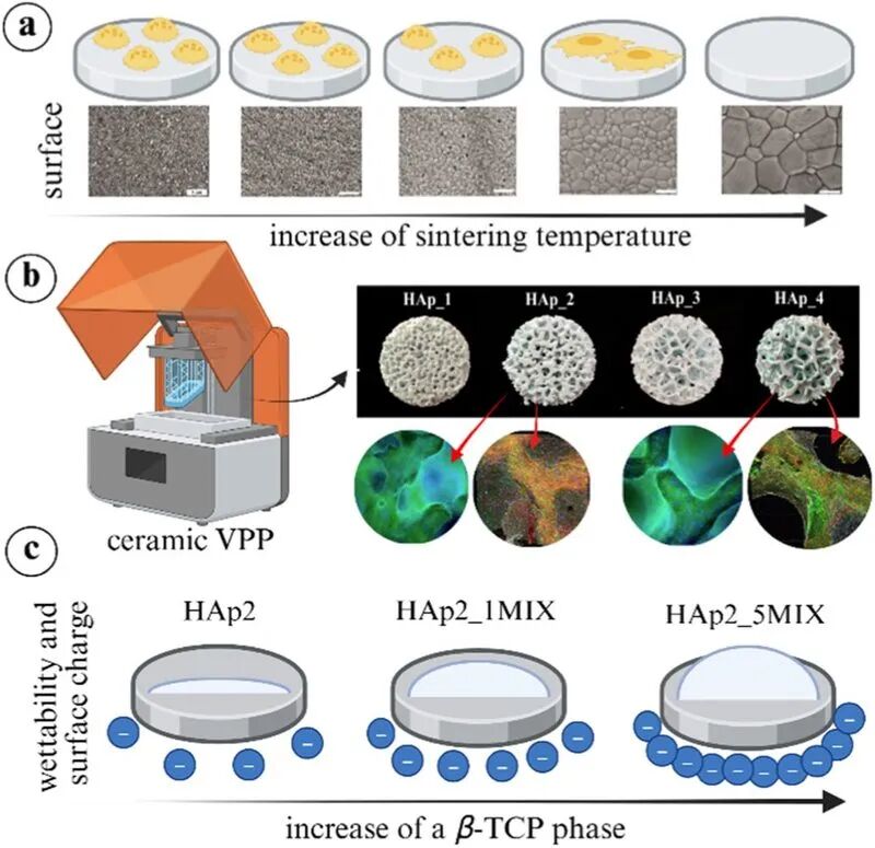
Using ceramic vat photopolymerization on a Lithoz CeraFab 7500, the study fabricated four HAp scaffold architectures and related microstructure to mechanical and biological performance. Within this comparison, approximately 45% porosity and 400-micrometre pores gave the preferred balance. The study also showed that trace-element substitution followed by high-temperature conversion of HAp to beta-TCP could worsen surface properties and reduce cell attachment.
The source draws two practical implications for this material system: sintering pure-HAp scaffolds at around 1000°C helps preserve bioactivity, and simply adding Sr, Mg or Zn ions to HAp slurry does not guarantee a benefit because subsequent phase transformation may reverse the intended effect. These are study-specific findings, not universal scaffold-design limits.
The authors identify bioceramic–polymer composites as a future direction: low-temperature processing may combine ceramic strength with polymer toughness while avoiding sintering-related changes and more closely approximating natural bone, which contains HAp and type I collagen.
References
[1] A. Ressler, R. Ohlsbom, V.A. Gobbo, M. Hannula, K. Keck, et al. Biomimetic bone calcium phosphate-based scaffolds fabricated via ceramic vat photopolymerization: Effect of porosity, sintering temperature, mineralogical phases and trace elements on the osteogenic potential. Materials Today Bio 38, 103140 (2026). DOI: 10.1016/j.mtbio.2026.103140.
[2] Y. Deng, D. Wang, W. Wang, et al. Effect of pore size and porosity on the mechanical properties and in vitro biological response of bioceramic scaffolds. Ceramics International, 2024. Reference as listed in the source.