Bioceramics
Reactive DLP: converting alpha-TCP to CDHA inside a bone scaffold
A printed photopolymer scaffold is hydrated at 37 °C to create an interpenetrating CDHA network while retaining substantial deformability.
Authorized republication with translated text and the source’s original scientific figures. Numerical results belong to the reported study, not to KUNWU equipment specifications. This is laboratory or preclinical research, not a clinical recommendation or evidence of a medical device’s clinical efficacy.
- Conversion after seven days
- >92%
- BET for P40/CDHA
- 16.31 m2 g-1
- Dimensional recovery
- 92.6–98.5%
Conventional bone-repair scaffolds face a dilemma: fully ceramic sintered parts are brittle and can fracture during surgery, while tougher polymer–ceramic composites contain isolated ceramic particles with limited reinforcing efficiency. A team at the Universitat Politècnica de Catalunya (UPC) proposed reactive DLP: incorporating hydrolysable alpha-TCP into a resin, printing the scaffold and then immersing it in water. Within seven days, more than 92% of the alpha-TCP converted in situ to calcium-deficient hydroxyapatite (CDHA). Interpenetrating networks of needle-like CDHA nanocrystals almost doubled compressive strength and modulus while retaining approximately 10% fracture strain, suggesting a new print-and-harden route for bone tissue engineering.
1. The brittleness dilemma in DLP bone scaffolds
Bone tissue engineering scaffolds require biocompatibility, appropriate mechanical properties and interconnected pores. DLP ceramic 3D printing can produce complex integrated geometries at resolutions of tens of micrometres and has therefore attracted increasing attention. However, a material trade-off remains. Fully ceramic scaffolds require high-temperature sintering and resemble natural bone mineral chemically, but their brittleness makes press-fitting or screw fixation difficult. Polymer–ceramic composites improve toughness, yet their ceramic phase usually remains as isolated particles in the polymer matrix, limiting reinforcement.
An alternative is an interpenetrating composite in which both ceramic and polymer form continuous, intertwined three-dimensional networks. DIW had already demonstrated this concept: after printing alpha-TCP/PCL ink, alpha-TCP hydrolyses into CDHA and self-hardens into an interpenetrating structure within the strands. However, nozzle diameters greater than 100 micrometres limit DIW resolution, making natural bone’s multiscale hierarchy and complex curved biomimetic features difficult to reproduce.
2. Reactive resin design: the dual role of alpha-TCP
The formulations used aqueous polyethylene glycol diacrylate (PEGDA, molecular weight 700 g/mol), alpha-TCP powder, the photoinitiator LAP and light absorbers Tartrazine/Orange-G. Four PEGDA contents—40, 60, 80 and 100 wt.%—were designated P40, P60, P80 and P100.
Rheology is the first requirement for DLP printability. Alpha-TCP increased resin viscosity substantially, but adding 2 wt.% Disperbyk 111 dispersant reduced all formulations below 1 Pa·s. Herschel–Bulkley fitting showed shear-thinning behavior (n < 1) and near-zero yield stress, supporting smooth layer spreading.
Photocuring kinetics produced an unexpected result: real-time photorheology showed that alpha-TCP accelerated photocrosslinking. The initial delay fell from approximately 18 s to 10 s and the maximum curing rate increased. Contrary to the usual light-blocking effect of ceramic particles, the team suggested that rapid alpha-TCP hydration consumed free water and locally concentrated the polymer. FTIR measurements supported this interpretation: degree of conversion exceeded 95% for all groups except P80, which reached 82%.
An LCD-DLP printer operating at 405 nm produced gyroid triply periodic minimal-surface (TPMS) scaffolds with 50% designed porosity and an average pore size of approximately 628 micrometres, comparable to cancellous bone. Micro-CT and SEM confirmed good interlayer bonding without obvious defects. Adding the reactive ceramic powder therefore preserved printability.
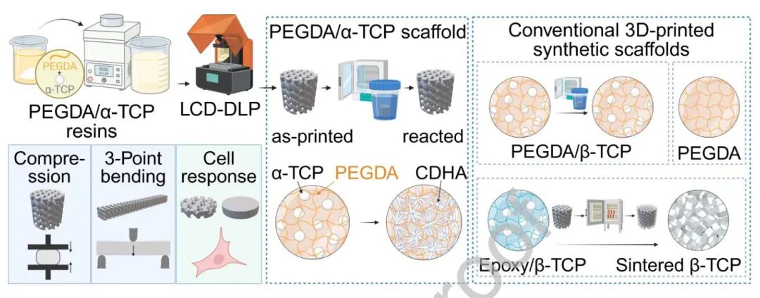
3. Seven days in water: in situ conversion from alpha-TCP to CDHA
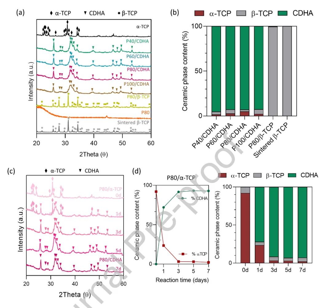
Alpha-TCP began hydrolysing as soon as the printed PEGDA/alpha-TCP scaffolds were immersed in water at 37°C. XRD showed approximately 72% conversion to CDHA on day 1. By day 7, conversion reached 95.1%, 92.7%, 92.8% and 92.5% for P40/CDHA, P60/CDHA, P80/CDHA and P100/CDHA, respectively, leaving only traces of beta-TCP and unreacted alpha-TCP. Rietveld quantitative phase analysis agreed with the diffraction changes.
SEM revealed bundles of intertwined needle-like CDHA nanocrystals forming a three-dimensional network. Higher polymer content increased coverage of the crystals by PEGDA. The BET specific surface area of P40/CDHA reached 16.31 m2/g, far above pure P80 polymer at 0.08 m2/g and sintered beta-TCP at 0.37 m2/g. This nanoscale roughness provides surface cues relevant to osteoinduction and bone formation.
4. Almost doubled mechanical properties: the benefit of interpenetrating networks
Mechanical tests compared the materials before and after reaction in the wet state to approximate physiological conditions. As-printed PEGDA/alpha-TCP was polymer-dominated, flexible and had approximately 15–20% fracture strain. After seven days of hydration, the CDHA network carried load: compressive strength and elastic modulus almost doubled while fracture strain remained around 10%. Strain-energy density, a combined toughness measure, also nearly doubled.
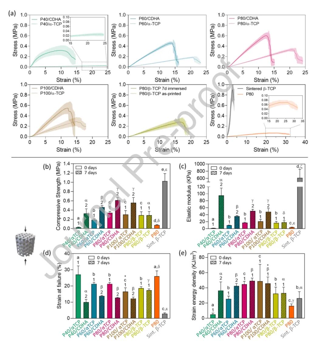
Three-point bending further highlighted the composite’s potential: P80/CDHA had more than 16 times the fracture displacement and five times the bending strain-energy density of the sintered beta-TCP control. A non-reactive P80/beta-TCP control showed no mechanical change after water immersion, ruling out swelling as the cause and linking reinforcement to CDHA formation.
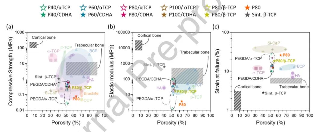
Compared with published DLP calcium-phosphate composites—including hydroxyapatite, silicon-substituted calcium phosphate and biphasic calcium phosphate—the PEGDA/CDHA scaffolds had comparable compressive strength and modulus but markedly greater fracture strain. Even after conversion, they retained substantial deformability. The team associated this behavior with the continuous concave surfaces of the gyroid lattice, which sacrifice some axial load-bearing capacity in exchange for a biological environment more favorable to osteoconduction.
5. Dry storage and water-triggered hardening: translational potential
Dimensional-stability measurements showed that swollen scaffolds could be freeze-dried for storage and recover 92.6–98.5% of their dimensions after rehydration. The source proposes dry implantation: shaping during surgery, followed by swelling in body fluid and in situ alpha-TCP-to-CDHA hardening. This print-and-harden concept seeks to avoid the intraoperative fragility of conventional ceramic parts.
Biocompatibility was evaluated using MC3T3-E1 osteoblast-like cells, with both indirect extract-based cytotoxicity testing (the source cites ISO 10993:12) and direct-contact experiments. LDH measurements showed no obvious cytotoxicity for any PEGDA/CDHA formulation, while confocal microscopy showed cell adhesion and spreading. A 28-day stability test in PBS also showed controlled mass changes, providing preliminary—not clinical—evidence for longer-term use.
References
Fagotto-Clavijo R, Lodoso-Torrecilla I, Diez-Escudero A, Ginebra M-P. Digital Light Processing of Reactive Ceramic-Polymer Bone Scaffolds Enables In situ Hydroxyapatite Formation. Acta Biomaterialia. 2026. DOI: 10.1016/j.actbio.2026.09.013.