Research applications
Zirconia TPMS scaffolds: combining strength, antibacterial activity and osteogenic function
A sequential research route: print a load-bearing zirconia skeleton, add a magnesium-doped calcium-silicate coating, then introduce alginate-protected gallium. All biological evidence remains in vitro.
Editorial translation of the supplied research article. Values describe the cited study, not guaranteed printer performance or a qualified KUNWU product. Original figure panels, labels and scale bars are unchanged. Publisher full text was not independently accessible during this update; check the original paper before reproducing its procedure.
1 · Structure first, function second
Bone-repair scaffolds need mechanical support, resistance to bacterial colonization and an environment that supports cell adhesion and osteogenic differentiation. Zirconia offers strength and chemical stability, but is biologically inert and not inherently antibacterial. The supplied study by teams at Hangzhou Dianzi University, Zhejiang Chinese Medical University and Zhejiang University combines three sequential operations rather than claiming that printing alone supplies all functions.
A high-solids zirconia framework is formed by projection vat photopolymerization and sintered. Vacuum adsorption then adds bioactive magnesium-doped calcium silicate (CSi-Mg), and sodium-alginate-protected gallium nanoparticles occupy the pores. Gyroid TPMS distributes load through continuous curved surfaces while retaining interconnected space for tissue ingrowth. The source contrasts this with weaker degradable scaffolds, bioinert zirconia and earlier calcium-silicate or hydroxyapatite coatings. Gallium is intended to interfere with bacterial iron metabolism; alginate protects the liquid-metal particles and helps immobilize them.
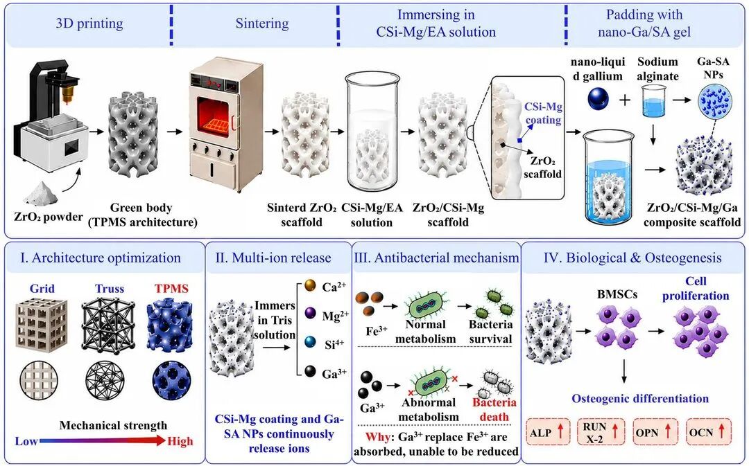
2 · Materials and printing
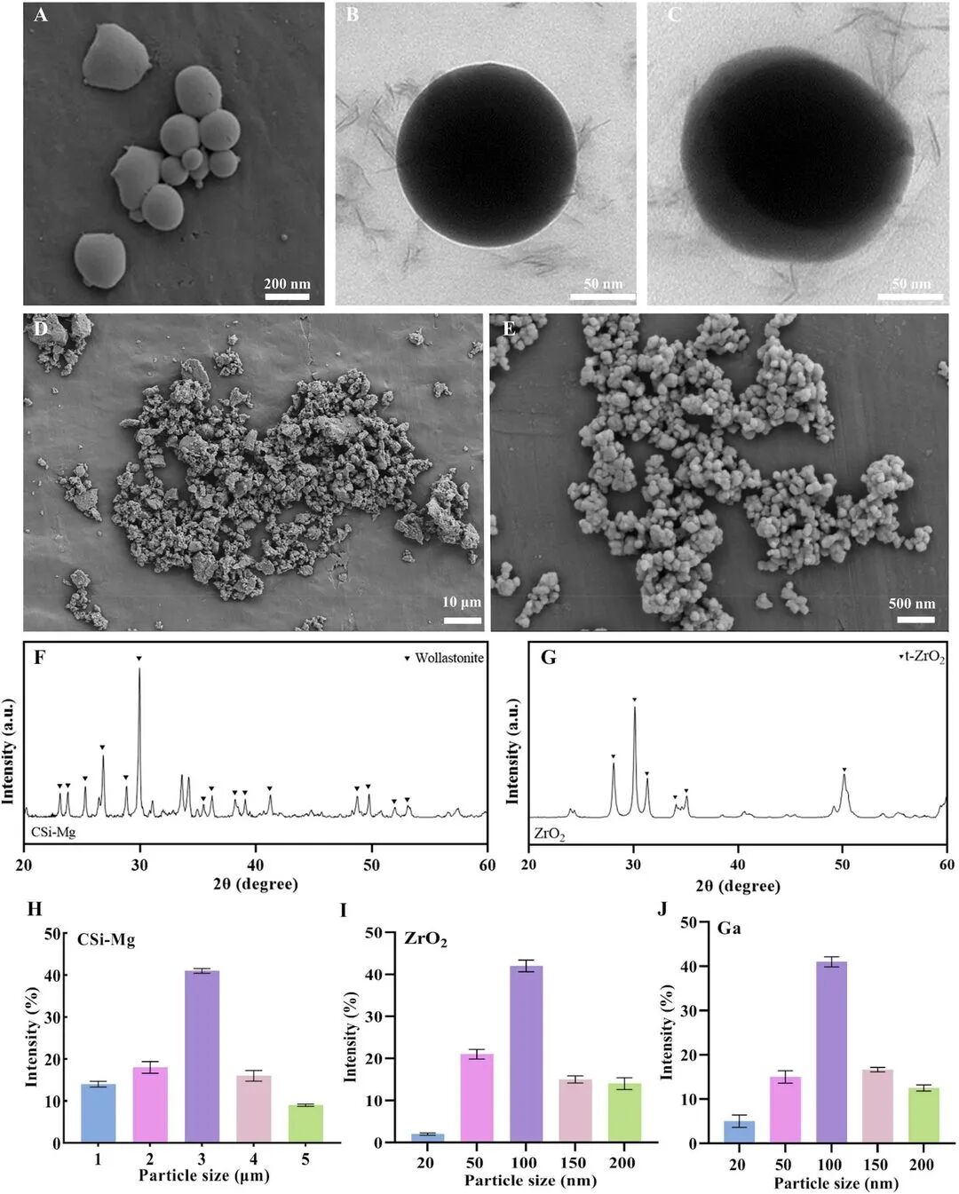
The zirconia is yttria-stabilized tetragonal powder, typically 50–150 nm. Wet-chemically precipitated CSi-Mg matches β-wollastonite (PDF 27-0088). Gallium is dispersed in 2 wt% aqueous sodium alginate by ice-bath ultrasonication and centrifugation, producing approximately 50–200 nm spherical Ga-SA particles. SEM, TEM, XRD and size distributions characterize the three precursors.
Printing slurries use HDDA/TMPTA monomers at 8:3, photoinitiator at 2 wt% relative to resin and dispersant at 2 wt% relative to powder. The source compares 70%, 75% and 80% solids loading. Printed scaffolds are cleaned, dried and sintered at 1500 °C. These are study-specific formulations, not a universal operating recipe for a printer.
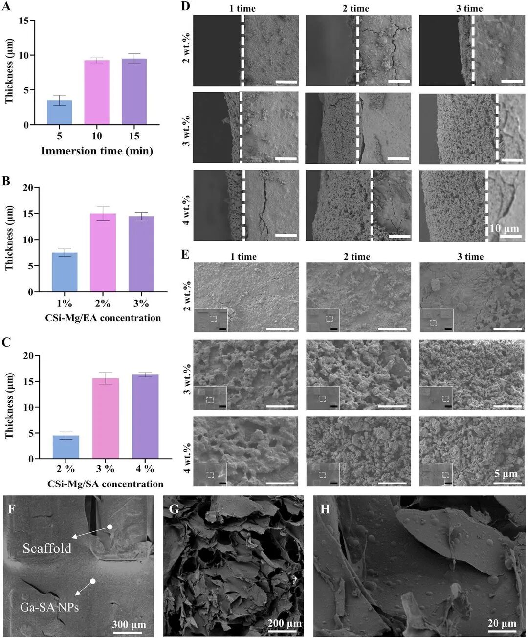
3 · Coating without blocking the pores
Coating thickness approaches a plateau after 10 minutes of impregnation; extending to 15 minutes offers little gain and risks blocking pores. At 10 minutes, 2% CSi-Mg/ethanol gives about 15 μm thickness; 3% does not increase it further. The CSi-Mg/alginate solution reaches its maximum at 3%. Alternating ethanol and alginate baths protects the previous layer and provides a substrate for the next adsorption cycle.
Three cycles of 10-minute vacuum impregnation followed by 20-minute drying at 80 °C produce ZrO₂/CSi-Mg. Dispersions containing 0%, 2% or 5% Ga-SA particles are then vacuum-infiltrated, calcium-chloride-crosslinked and freeze-dried. Cross-sections show gel attached along the internal channels and relatively uniform nanoparticle dispersion without obvious aggregates.
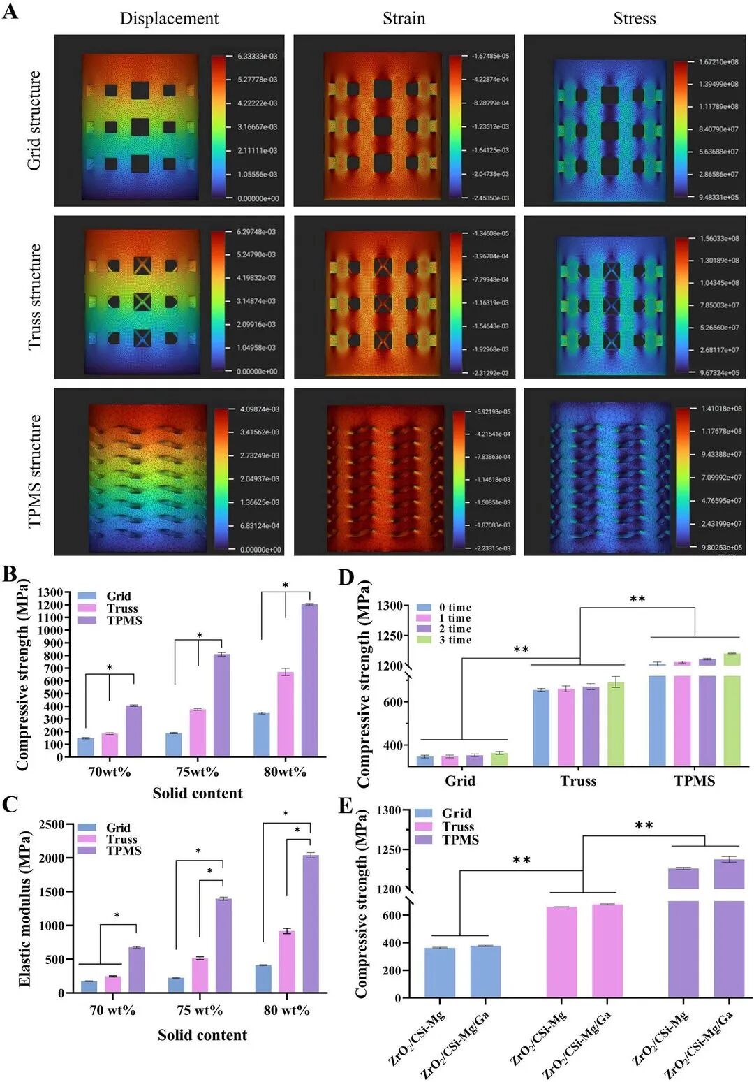
4 · Geometry and solids loading control strength
The source describes approximately 6 mm test specimens, about 32.5% porosity and 300 μm average pores, loaded along Z, perpendicular to print layers, at 0.5 mm/s. Under the same test conditions, raising solids from 70% to 80% increases compressive strength from 150 to 351 MPa for a grid, 189 to 650 MPa for a truss and 410 to 1200 MPa for Gyroid TPMS. The TPMS gain is 790 MPa versus 201 MPa for the grid. Modulus follows a similar ranking; simulations associate TPMS with more uniform stress transfer.
Neither CSi-Mg adsorption nor Ga-SA filling reduces the reported strength; both give modest increases, with coated values above 1230 MPa. These unusually high values belong to the reported geometry and test setup and must not be transferred to arbitrary porosity, specimen shape or clinical load cases. The source description of specimen shape and some percentage changes is internally ambiguous; consult the paper and raw figure before reproducing or specifying a test.
5 · Three-week in-vitro immersion
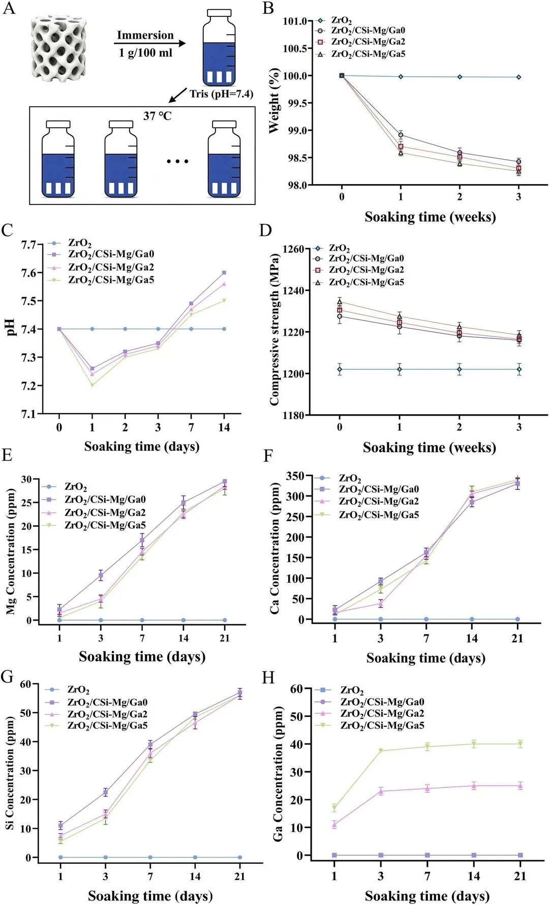
Scaffolds are immersed in Tris-HCl at pH 7.4 and 37 °C for 1–3 weeks. At three weeks, Ga0/Ga2/Ga5 lose approximately 1.5/1.6/1.7% mass, while unmodified zirconia loses almost none. Coated groups raise solution pH toward 7.6. The zirconia framework remains the mechanical support as the coating releases functional ions; modified groups retain approximately 1222 MPa in the source, above the unmodified group near 1200 MPa.
Ca, Mg, silicate and Ga release increase with immersion time. Ga2 and Ga5 have comparable Ca/Mg/Si release, but Ga5 releases more gallium. Limited coating degradation explains the small mass and strength changes; it does not establish in-vivo resorption or long-term implant stability.
6 · Antibacterial activity has a dosage window
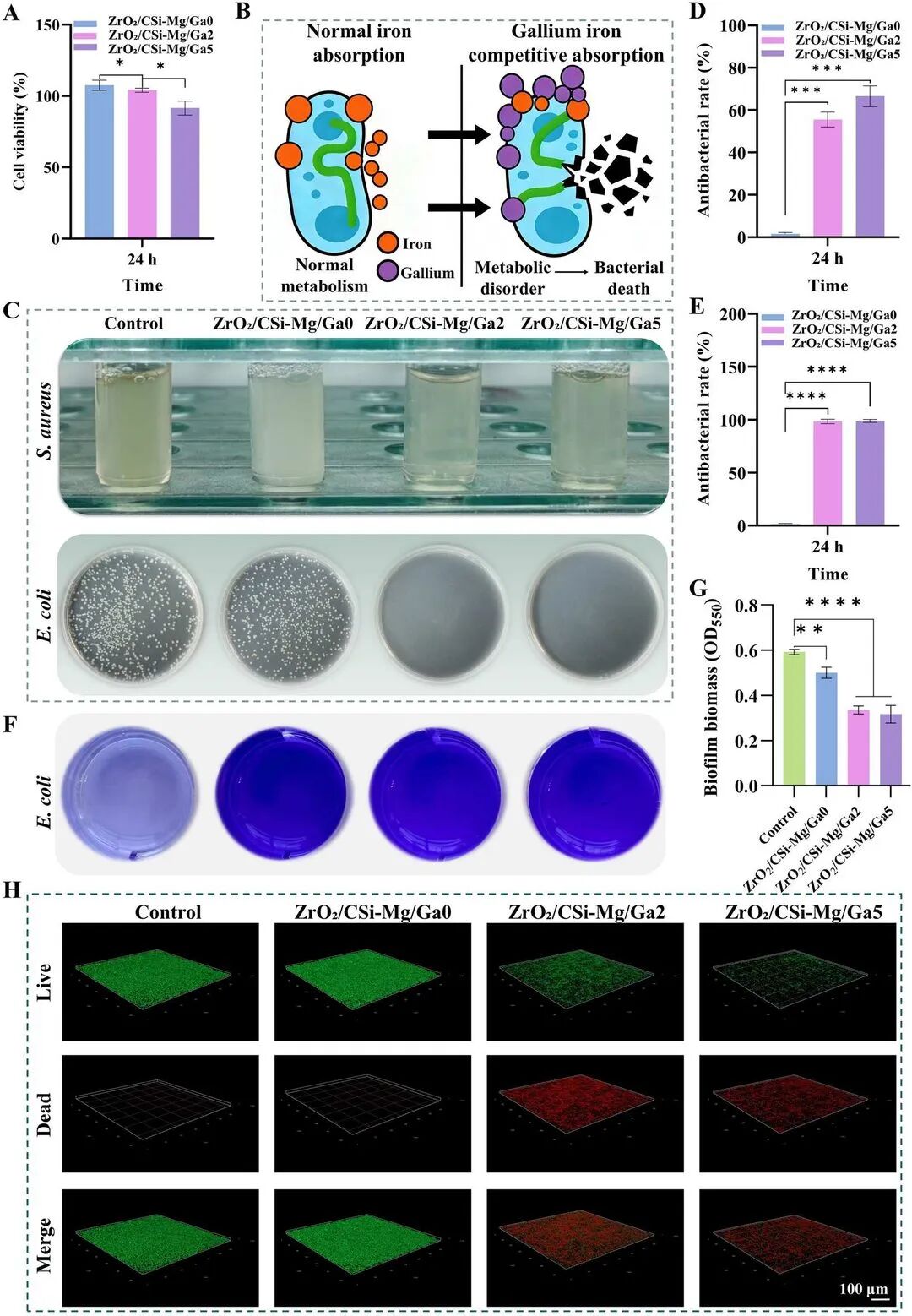
After 24-hour co-culture, Ga0, the coated but gallium-free control, has almost no activity against S. aureus and no clear E. coli effect. Ga2 and Ga5 show approximately 55% and 67% activity against S. aureus, respectively, and nearly 100% against E. coli. Crystal-violet biomass measurements and confocal live/dead biofilm imaging support the antibacterial effect. Gallium is proposed to replace Fe³⁺ in bacterial metabolism, disrupting normal function.
Higher gallium is not automatically better: Ga5 reduces cell activity over time, whereas Ga0 and Ga2 do not show the same inhibition. The authors therefore take Ga2 forward as the balance between antibacterial effect and cytocompatibility. This window must be calibrated for each material and biological system.
7 · Osteogenic evidence and its limits
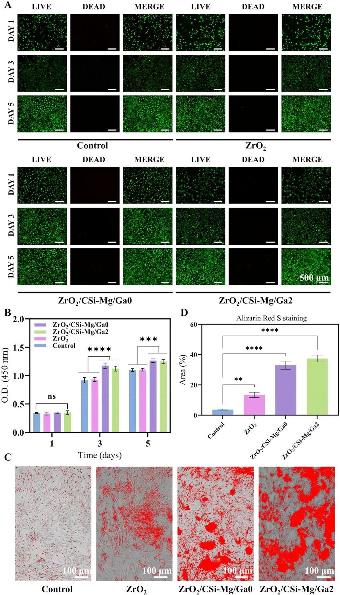
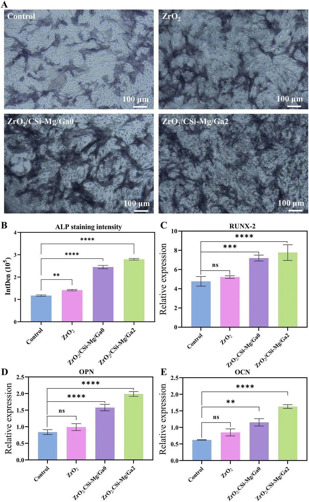
Bone-marrow mesenchymal stem cells are compared on pure zirconia, Ga0 and Ga2. Live/dead staining at days 1, 3 and 5 and CCK-8 show more cells on the coated groups; Ga0 has the highest activity, followed by Ga2. Alizarin-red staining shows greater calcium deposition in both coated groups and more in Ga2 than Ga0. Ga2 gives the strongest ALP staining and higher RUNX-2, OPN and OCN expression, spanning early, intermediate and late osteogenic markers. The source attributes the result to combined Ca/Mg/Si/Ga activity rather than zirconia alone.
The transferable lesson is the sequence: optimize scaffold geometry and solids loading, add bioactivity without closing pores, then tune antibacterial filling. All degradation, bacterial and cell tests reported here are in vitro. They do not demonstrate clinical safety, approved implantation or bone repair in a living patient. The paper calls for suitable in-vivo defect models before translation. KUNWU presents this as a materials-research example, not a medical treatment or implant offer.
Sources
Original paper · DOI
Xiao J, Tang X, Zhou C, et al. Multifunctional design of 3D-printed TPMS-structured ZrO₂/CSi-Mg/Ga composite scaffolds for enhanced mechanical, antibacterial, and osteogenic performance. Virtual and Physical Prototyping, 2026, 21(1): e2740292.